Showing posts with label FISIKA SINAR-X. Show all posts

Monday, 26 August 2013

FISIKA SINAR-X,


PENGENALAN SINAR-X 
1.     Pengertian Sinar-X
Sinar –X adalah gelombang elektromagnetik yang mempunyai panjang gelombang 10-8 -10-12 m dan frekuensi sekitar 1016 -1021 Hz. Sinar ini dpat menembus benda-benda lunak seperti daging dan kulit tetapi tidak dapat menembus benda-benda keras seperti tulang, gigi, dan logam. Sinar-X sering di gunakan di berbagai bidang seperti bidang kedokteran, fisika, kimia, mineralogy, metarulugi, dan biologi.
2.     Sejarah Penemuan Sinar-X
Sinar-X ditemukan pertama kali oleh fisikawan berkebangsaan Jerman Wilhelm C. Roentgen pada tanggal 8 November 1895. Saat itu Roentgen bekerja menggunakan tabung. Dia mengamati nyala hijau pada tabung yang sebelumnya menarik perhatian Crookes. Roentgen selanjutnya mencoba menutup tabung itu dengan kertas hitam dengan harapan agar tidak ada cahaya tampak yang dapat lewat. Namun setelah ditutup ternyata masih ada sesuatu yang dapat lewat. Roentgen Menyimpulkan bahwa ada sinar-sinar tidak tampak yang mampu menerobos kertas hitam tersebut.
Pada saat Roentgen menyalakan sumber listrik tabung untuk penelitian sinar katoda, beliau mendapatkan bahwa ada sejenis cahaya berpendar pada layar yang terbuat dari barium platino cyanida yang kebetulan berada di dekatnya. Jika sumber listrik dipadamkan, maka cahaya pendar pun hilang. Roentgen segera menyadari bahwa sejenis sinar yang tidak kelihatan telah muncul dari dalam tabung sinar katoda. Karena sebelumnya tidak pernah dikenal, maka sinar ini diberi nama sinar-X. Namun untuk menghargai jasa beliau dalam penemuan ini maka seringkali sinar-X itu dinamai juga sinar Roentgen.
            Nyala hijau yang terlihat oleh Crookes dan Roentgen akhirnya diketahui bahwa sinar tersebut tak lain adalah gelombang cahaya yang dipancarkan oleh dinding kaca pada tabung sewaktu elektron menabrak dinding itu, sebagai akibat terjadinya pelucutan listrik melalui gas yang masih tersisa di dalam tabung. Pada saat yang bersamaan elektron itu merangsang atom pada kaca untuk mengeluarkan gelombang elektromagnetik yang panjang gelombangnya sangat pendek dalam bentuk sinar-X. Sejak saat itu para ahli fisika telah mengetahui bahwa sinar-X dapat dihasilkan bila elektron dengan kecepatan yang sangat tinggi menabrak atom.
            Tergiur oleh penemuannya yang tidak sengaja itu, Roentgen memusatkan perhatiannya pada penyelidikan sinar-X. Dari penyelidikan itu beliau mendapatkan bahwa sinar-X dapat memendarkan berbagai jenis bahan kimia. Sinar-X juga dapat menembus berbagai materi yang tidak dapat ditembus oleh sinar tampak biasa yang sudah dikenal pada saat itu. Di samping itu, Roentgen juga bisa melihat bayangan tulang tangannya pada layar yang berpendar dengan cara menempatkan tangannya di antara tabung sinar katoda dan layar. Dari hasil penyelidikan berikutnya diketahui bahwa sinar-X ini merambat menempuh perjalanan lurus dan tidak dibelokkan baik oleh medan listrik maupun medan magnet. Atas jasa-jasa Roentgen dalam menemukan dan mempelajari sinar-X ini, maka pada tahun 1901 beliau dianugerahi Hadiah Nobel Bidang Fisika yang untuk pertama kalinya diberikan dalam bidang ini. Penemuan Sinar-X ternyata mampu mengantarkan ke arah terjadinya perubahan mendasar dalam bidang kedokteran. Dalam kegiatan medis, Sinar-X dapat dimanfaatkan untuk diagnosa maupun terapi. Dengan penemuan sinar-X ini, informasi mengenai tubuh manusia menjadi mudah diperoleh tanpa perlu melakukan operasi bedah.
            Sinar-X dapat terbentuk apabila partikel bermuatan misalnya elektron oleh pengaruh gaya inti atom bahan mengalami perlambatan. Sinar-X yang tidak lain adalah gelombang elektromagnetik yang terbentuk melalui proses ini disebut sinar-X bremsstrahlung. Sinar-X yang terbentuk dengan cara demikian mempunyai energi paling tinggi sama dengan energi kinetik partikel bermuatan pada waktu terjadinya perlambatan.
 SinarX-Bremstrahlung 
Gambar. Eksitasi elektron sinar-X
Ketika terjadi perlambatan dan menimbulkan sinar-X, sinar-X yang terjadi umumnya memiliki energi yang berbeda-beda sesuai dengan energi kinetik elektron pada saat terbentuknya sinar-X dan juga bergantung pada arah pancarannya.
                        x-ray
                        Gambar. Proses pembentukan sinar-X memakai tabung katoda-anoda
            Sinar-X dapat juga terbentuk dalam proses perpindahan elektron-elektron atom dari tingkat energi yang lebih tinggi menuju ke tingkat energi yang lebih rendah, misalnya dalam proses lanjutan efek fotolistrik. Sinar-X yang terbentuk dengan cara seperti ini mempunyai energi yang sama dengan selisih energi antara kedua tingkat energi yang berkaitan. Karena energi ini khas untuk setiap jenis atom, sinar yang terbentuk dalam proses ini disebut sinar-X karakteristik, kelompok sinar-X demikian mempunyai energi farik. Sinar-X karakteristik yang timbul oleh berpindahnya elektron dari suatu tingkat energi menuju ke lintasan k, disebut sinar-X garis K, sedangkan yang menuju ke lintasan l, dan seterusnya. Sinar-X bremsstrahlung dapat dihasilkan melalui pesawat sinar-X atau pemercepat partikel.
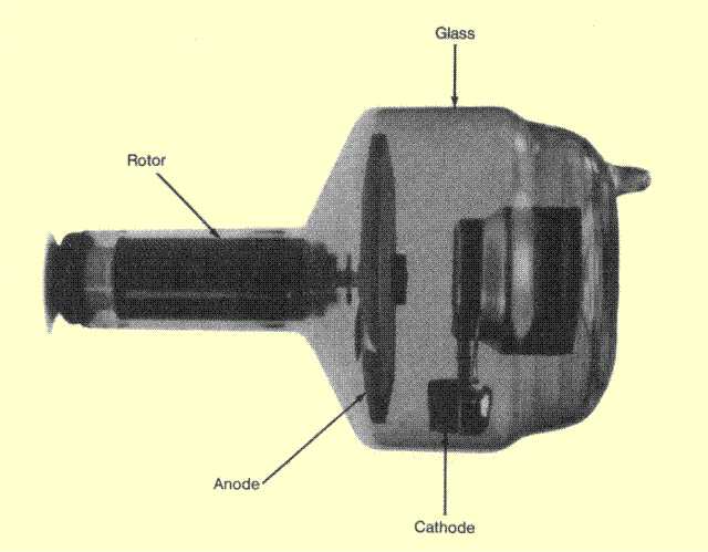
            Pada dasarnya pesawat sinar-X terdiri dari tiga bagian utama, yaitu tabung sinar-X, sumber tegangan tinggi yang mencatu tegangan listrik pada kedua elektrode dalam tabung sinar-X, dan unit pengatur. Bagian pesawat sinar-X yang menjadi sumber radiasi adalah tabung sinar-X. Didalam tabung pesawat sinar-X yang biasanya terbuat dari bahan gelas terdapat filamen yang bertindak sebagai katode dan target yang bertindak sebagai anode. Tabung pesawat sinar-X dibuat hampa udara agar elektron yang berasal dari filamen tidak terhalang oleh molekul udara dalam perjalanannya menuju ke anode. Filamen yang di panasi oleh arus listrik bertegangan rendah (If) menjadi sumber elektron. Makin besar arus filamen IF, akan makin tinggi suhu filamen dan berakibat makin banyak elektron dibebaskan persatuan waktu.

            Elektron yang dibebaskan oleh filamen tertarik ke anode oleh adanya beda potensial yang besar atau tegangan tinggi antara katode dan anode yang dicatu oleh unit sumber tegangan tinggi (potensial katode beberapa puluh hingga beberapa ratus kV atau MV lebih rendah dibandingkan potensial anode), elektron ini menabrak bahan target yang umumnya bernomor atom dan bertitik cair tinggi (misalnya tungsten) dan terjadilah proses bremsstrahlung. Khusus pada pemercepat partikel energi tinggi beberapa elektron atau partikel yang dipercepat dapat agak menyimpang dan menabrak dinding sehingga
menimbulkan bremsstrahlung pada dinding. Beda potensial atau tegangan antara kedua elektrode menentukan energi maksimum sinar-X yang terbentuk, sedangkan fluks sinar-X bergantung pada jumlah elektron persatuan waktu yang sampai ke bidang anode yang terakhir ini disebut arus tabung It yang sudah barang tentu bergantung pada arus filamen It. Namun demikian dalam batas tertentu, tegangan tabung juga dapat mempengaruhi arus tabung. Arus tabung dalam sistem pesawat sinar-X biasanya hanya mempunyai tingkat besaran dalam milliampere (mA), berbeda dengan arus filamen yang besarnya dalam tingkat ampere.
Wilhelm_Roentgen  Gambar: Penemu sinar-X
3.     Cara Kerja Sinar-X
            Pada aplikasinya, penciptaan sinar-x tak lagi mengandalkan mekanisme tabung crookes, melakinkan dengan menggunakan pesawat sinar-x modern. Pesawat sinar-x modern pada dasarnya membangkitkan sinar-x dengan mem’bombardir’ target logam dengan elektron berkecepatan tinggi. Elektron yang berkecepatan tinggi tentunya memiliki energi yang tinggi, dan karenanya mampu menembus elektron-elektron orbital luar pada materi target hingga menumbuk elektron orbital pada kulit k (terdekat dengan inti).
            Elektron yang tertumbuk akan terpental dari orbitnya, meninggalkan hole pada tempatnya semula. Hole yang ditinggalkannya itu akan diisi oleh elektron dari kulit luar dan proses itu melibatkan pelepasan foton (cahaya elektromagnetik) dari elektron pengisi tersebut. Foton yang keluar itulah yang kemudian disebut sinar-x, dan keseluruhan proses terbentuknya sinar-x melalui mekanisme tersebut disebut mekanisme sinar-x karakteristik.
            Adapun mekanisme lain yang mungkin terjadi adalah emisi foton yang dialami oleh elektron cepat yang dibelokkan oleh inti atom target atas konsekuensi dari interaksi coulomb antara inti atom target dengan elektron cepat. Proses pembelokkan ini melibatkan perlambatan dan karenanya memerlukan emisi energi berupa foton. Mekanisme ini disebut bremsstrahlung (bahasa jerman dari ‘radiasi pengereman’).
2    
Gambar. Proses produksi sinar-X
            Seperti terlihat pada gambar ilustrasi, beda potensial antara anoda dan katoda dibuat sedemikian rupa sehingga mencapai angka yang cukup untuk membuat elektron melompat dengan kecepatan tinggi setelah katoda diberi energy (biasanya 1000 volt). Setelah elektron pada katoda melompat dan menghantam filamen pada anoda, terjadilah sinar-x yang terjadi dengan mekanisme sinar-x karakteristik ataupun bremsstrahlung. Karena filamen pada anoda dimiringkan ke bawah, foton sinar-x akan menuju ke bawah, keluar dari pesawat sinar-x lalu melewati jaringan yang dipotret. Bayangan/citra pun terbentuk pada film yang diletakkan di bawahnya.

Aplikasi Sinar-X Dalam Kehidupan Sehari-Hari

1.     Bidang Kesehatan
·        Sinar-X energirendah digunakan untuk mengambil gambar foto yang dikenal sebagai radiograf. Sinar-X bisa menembus tubuh manusia tetapi diserap oleh bagian yang lebih padat seperti tulang.
3  
Gambar foto sinar-X digunakan untuk memperlihatkan kecacatan tulang, mengdeteksi tulang yang patah dan memperlihatkan keadaan organ-organ dalam tubuh.

·        Sinar-X keras digunakan untuk memusnahkan sel-sel kanker. Cara ini dikenal sebagai radioterapi.
2.     Dalam Bidang Perindustrian
·      Mengetahui kecacatan dalam struktur binaan atau bagian-bagian dalam mesin dan engine.
·      Memperbaiki rekahan dalam pipa logam, dinding konkrit dan tekanan tinggi.
·      Memeriksa retakan dalam struktur plastik dan getah.
3.     Dalam Bidang Penyelidikan
·        Sinar-X digunakan untuk menyelidik struktur hablur dan jarak pemisahan antara atom-atom dalam suatu bahan hablur.
4.     Kegunaan lainnya
·        Sinar-X digunakan untuk mengesahkan sama ada suatu lukisan atau objek seni purba itu benar atau tiruan.
·        Di lapangan kapal terbang, sinar-X lembut digunakan untuk memeriksa barang-barang dan beg penumpang.
Efek Yang Ditimbulkan Dari Sinar-X
            Efek merugikan tersebut berupa kerontokan rambut dan kerusakan kulit. Pada tahun 1897 di Amerika Serikat dilaporkan adanya 69 kasus kerusakan kulit yang disebabkan oleh sinar-X, sedang pada tahun 1902 angka yang dilaporkan meningkat menjadi 170 kasus. Pada tahun 1911 di Jerman juga dilaporkan adanya 94 kasus tumor yang disebabkan oleh sinar-X. Meskipun beberapa efek merugikan dari sinar-X dan gamma telah teramati, namun upaya perlindungan terhadap bahaya penyinaran sinar-X dan gamma belum terfikirkan. Marie Curie, penemu bahan radioaktif Po dan Ra meninggal pada tahun 1934 akibat terserang oleh leukemia. Penyakit tersebut besar kemungkinan akibat paparan radiasi karena seringnya beliau berhubungan dengan bahan-bahan radioaktif.
Efek lainnya yaitu :
·        Pemusnahan sel-sel dalam tubuh.
·        Perubahan struktur genetik suatu sel.
·        Penyakit kanser darah.
·         Kesan-kesan buruk seperti rambut rontok, kulit menjadi merah dan berbisul
http://hadirwong.blogspot.com/2009/12/manfaat-sinar-x.html


Tuesday, 12 June 2012

X-Ray ProductionPerry Sprawls, Ph.D.
Online Textbook

INTRODUCTION AND OVERVIEW
THE X-RAY TUBE
   Function
   Anode
   Design
   Focal Spot
   Cathode
   Envelope
   Housing
THE X-RAY CIRCUIT
ELECTRON ENERGY
   Potential
   Kinetic
   Binding
BREMSSTRAHLUNG
   Production Process
   Spectrum
   Effect of KV
CHARACTERISTIC RADIATION
    Production
   Tungsten Spectrum
   Molybdenum Spectrum
   Rhodium Spectrum
   KV Effect on Spectrum
EFFICIENCY
   Concept
   Effect of KV
   Anode Material
EFFICACY (OUTPUT)
   Definition and Concept
   KV Control
   Waveform
SUMMARY and MIND MAP

INTRODUCTION AND OVERVIEW

  X-radiation is created by taking energy from electrons and converting it into photons with appropriate energies. This energy conversion takes place within the x-ray tube. The quantity (exposure) and quality (spectrum) of the x-radiation produced can be controlled by adjusting the electrical quantities (KV, MA) and exposure time, S, applied to the tube. In this chapter we first become familiar with the design and construction of x-ray tubes, then look at the x-ray production process, and conclude by reviewing the quantitative aspects of x-ray production.

   Function

    An x-ray tube is an energy converter. It receives electrical energy and converts it into two other forms: x-radiation and heat. The heat is an undesirable byproduct. X-ray tubes are designed and constructed to maximize x-ray production and to dissipate heat as rapidly as possible.
   The x-ray tube is a relatively simple electrical device typically containing two principle elements: a cathode and an anode. As the electrical current flows through the tube from cathode to anode, the electrons undergo an energy loss, which results in the generation of x-radiation. A cross-sectional view of a typical x-ray tube is shown in below.

Cross-Section of a Typical X-Ray Tube

   Anode

  The anode is the component in which the x-radiation is produced. It is a relatively large piece of metal that connects to the positive side of the electrical circuit.
   The anode has two primary functions: (1) to convert electronic energy into x-radiation, and (2) to dissipate the heat created in the process. The material for the anode is selected to enhance these functions.
   The ideal situation would be if most of the electrons created x-ray photons rather than heat. The fraction of the total electronic energy that is converted into x-radiation (efficiency) depends on two factors: the atomic number (Z) of the anode material and the energy of the electrons. Most x-ray tubes use tungsten, which has an atomic number of 74, as the anode material. In addition to a high atomic number, tungsten has several other characteristics that make it suited for this purpose. Tungsten is almost unique in its ability to maintain its strength at high temperatures, and it has a high melting point and a relatively low rate of evaporation. For many years, pure tungsten was used as the anode material. In recent years an alloy of tungsten and rhenium has been used as the target material but only for the surface of some anodes. The anode body under the tungsten-rhenium surface on many tubes is manufactured from a material that is relatively light and has good heat storage capability. Two such materials are molybdenum and graphite. The use of molybdenum as an anode base material should not be confused with its use as an anode surface material. Most x-ray tubes used for mammography have molybdenum-surface anodes. This material has an intermediate atomic number (Z = 42), which produces characteristic x-ray photons with energies well suited to this particular application. Some mammography tubes also have a second anode made of rhodium, which has an atomic number of 45. This produces a higher energy and more penetrating radiation, which can be used to image dense breast.
   The use of a rhenium-tungsten alloy improves the long-term radiation output of tubes. With x-ray tubes with pure tungsten anodes, radiation output is reduced with usage because of thermal damage to the surface


     Design

   Most anodes are shaped as beveled disks and attached to the shaft of an electric motor that rotates them at relatively high speeds during the x-ray production process. The purpose of anode rotation is to dissipate heat and is considered in detail in another chapter.


 Focal Spot

   Not all of the anode is involved in x-ray production. The radiation is produced in a very small area on the surface of the anode known as the focal spot. The dimensions of the focal spot are determined by the dimensions of the electron beam arriving from the cathode. In most x-ray tubes, the focal spot is approximately rectangular. The dimensions of focal spots usually range from 0.1 mm to 2 mm. X-ray tubes are designed to have specific focal spot sizes; small focal spots produce less blurring and better visibility of detail, and large focal spots have a greater heat-dissipating capacity.
   Focal spot size is one factor that must be considered when selecting an x-ray tube for a specific application. Tubes with small focal spots are used when high image visibility of detail is essential and the amount of radiation needed is relatively low because of small and thin body regions as in mammography.
Most x-ray tubes have two focal spot sizes (small and large), which can be selected by the operator according to the imaging procedure. 


    Cathode

   The basic function of the cathode is to expel the electrons from the electrical circuit and focus them into a well-defined beam aimed at the anode. The typical cathode consists of a small coil of wire (a filament) recessed within a cup-shaped region, as shown below.


Energy Exchange within an X-Ray Tube

   Electrons that flow through electrical circuits cannot generally escape from the conductor material and move into free space. They can, however, if they are given sufficient energy. In a process known as thermionic emission, thermal energy (or heat) is used to expel the electrons from the cathode. The filament of the cathode is heated in the same way as a light bulb filament by passing a current through it. This heating current is not the same as the current flowing through the x-ray tube (the MA) that produces the x-radiation. During tube operation, the cathode is heated to a glowing temperature, and the heat energy expels some of the electrons from the cathode.


   Envelope

    The anode and cathode are contained in an airtight enclosure, or envelope. The envelope and its contents are often referred to as the tube insert, which is the part of the tube that has a limited lifetime and can be replaced within the housing. The majority of x-ray tubes have glass envelopes, although tubes for some applications have metal and ceramic envelopes.
   The primary functions of the envelope are to provide support and electrical insulation for the anode and cathode assemblies and to maintain a vacuum in the tube. The presence of gases in the x-ray tube would allow electricity to flow through the tube freely, rather than only in the electron beam. This would interfere with x-ray production and possibly damage the circuit.

   Housing

    The x-ray tube housing provides several functions in addition to enclosing and supporting the other components. It functions as a shield and absorbs radiation, except for the radiation that passes through the window as the useful x-ray beam. Its relatively large exterior surface dissipates most of the heat created within the tube. The space between the housing and insert is filled with oil, which provides electrical insulation and transfers heat from the insert to the housing surface.

THE X-RAY CIRCUIT

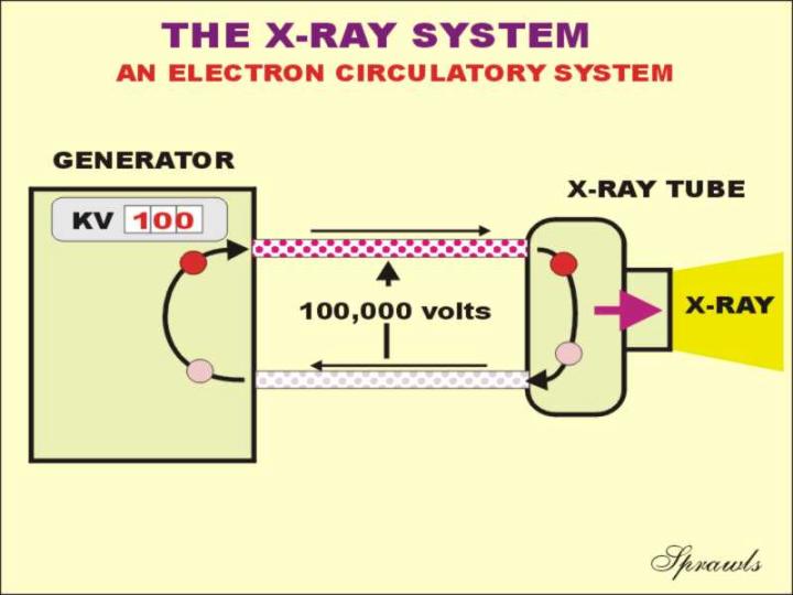
The energy used by the x-ray tube to produce x-radiation is supplied by an electrical circuit as illustrated
below.  The circuit connects the tube to the source of electrical energy, that in the x-ray room is often referred to as the
generator.  As described in another chapter, the generator receives the electrical energy from the electrical power system and converts it into the appropriate form (DC, direct current) to apply to the x-ray tube.  The generator also provides the ability to adjust certain electrical quantities that control the x-ray production process.
The three principle electrical quantities that can be adjusted are the:
  • KV (the voltage or electrical potential applied to the tube)
  • MA (the electrical current that flows through the tube)
  • S (duration of the exposure or exposure time, generally a fraction of a second)
The circuit is actually a circulatory system for electrons.  They pickup energy as the pass through the generator and transfer their energy to the x-ray tube anode as described above.


ELECTRON ENERGY

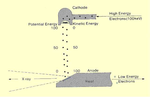
    The energy that will be converted into x-radiation (and heat) is carried to the x-ray tube by a current of flowing electrons as shown above. As the electrons pass through the x-ray tube, they undergo two energy conversions, as illustrated previously: The electrical potential energy is converted into kinetic (motion) energy that is, in turn, converted into x-radiation and heat.
 

   Potential

     When the electrons arrive at the x-ray tube, they carry electrical potential energy. The amount of energy carried by each electron is determined by the voltage or KV, between the anode and cathode. For each kV of voltage, each electron has 1 keV of energy. By adjusting the KV, the x-ray machine operator actually assigns a specific amount of energy to each electron.
 

   Kinetic

    After the electrons are emitted from the cathode, they come under the influence of an electrical force pulling them toward the anode. This force accelerates them, causing an increase in velocity and kinetic energy. This increase in kinetic energy continues as the electrons travel from the cathode to the anode. As the electron moves from cathode to anode, however, its electrical potential energy decreases as it is converted into kinetic energy all along the way. Just as the electron arrives at the surface of the anode its potential energy is lost, and all its energy is kinetic. At this point the electron is traveling with a relatively high velocity determined by its actual energy content. A 100-keV electron reaches the anode surface traveling at more than one half the velocity of light. When the electrons strike the surface of the anode, they are slowed very quickly and lose their kinetic energy; the kinetic energy is converted into either x-radiation or heat.    The electrons interact with individual atoms of the anode material, as shown below. Two types of interactions produce radiation. An interaction with electron shells produces characteristic x-ray photons; interactions with the atomic nucleus produce Bremsstrahlung x-ray photons.

Electron-Atom Interactions That Produce X-Ray Photon


   Binding Energy

The electrons within an atom each have a specific amount of binding energy that depends on the size (atomic number, Z) of the atom and the shell in which the electron is located.  As described in a previous chapter the binding energy is the energy that would be required to remove the electron from the atom.  It is actually an energy deficit rather than an amount of available energy.
The binding energy of electrons within an atom plays a major role in the production of characteristic x-radiation as described later.
 

   Production Process

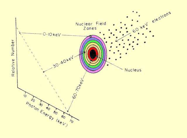
     The interaction that produces the most photons is the Bremsstrahlung process. Bremsstrahlung is a German word for "braking radiation" and is a good description of the process. Electrons that penetrate the anode material and pass close to a nucleus are deflected and slowed down by the attractive force from the nucleus. The energy lost by the electron during this encounter appears in the form of an x-ray photon. All electrons do not produce photons of the same energy


    Spectrum

    Only a few photons that have energies close to that of the electrons are produced; most have lower energies. Although the reason for this is complex, a simplified model of the Bremsstrahlung interaction is shown below. First, assume that there is a space, or field, surrounding the nucleus in which electrons experience the "braking" force. This field can be divided into zones, as illustrated. This gives the nuclear field the appearance of a target with the actual nucleus located in the center. An electron striking anywhere within the target experiences some braking action and produces an x-ray photon. Those electrons striking nearest the center are subjected to the greatest force and, therefore, lose the most energy and produce the highest energy photons. The electrons hitting in the outer zones experience weaker interactions and produce lower energy photons. Although the zones have essentially the same width, they have different areas. The area of a given zone depends on its distance from the nucleus. Since the number of electrons hitting a given zone depends on the total area within the zone, it is obvious that the outer zones capture more electrons and create more photons. From this model, an x-ray energy spectrum, such as the one shown below, could be predicted.


A Model for Bremsstrahlung Production and the Associated Photon Energy Spectrum

   The basic Bremsstrahlung spectrum has a maximum photon energy that corresponds to the energy of the incident electrons. This is 70 keV for the example shown. Below this point, the number of photons produced increases as photon energy decreases. The spectrum of x-rays emerging from the tube generally looks quite different from the one shown here because of selective absorption within the filter.
   A significant number of the lower-energy photons are absorbed or filtered out as they attempt to pass through the anode surface, x-ray tube window, or added filter material. X-ray beam filtration is discussed more extensively in a later chapter. The amount of filtration is generally dependent on the composition and thickness of material through which the x-ray beam passes and is generally what determines the shape of the low-energy end of the spectrum curve.


 Effect of KV

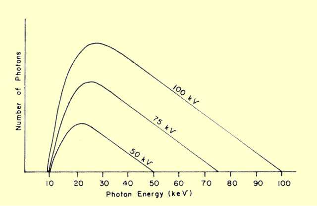
   The high-energy end of the spectrum is determined by the KV (kilovoltage) applied to the x-ray tube. This is because the KV establishes the energy of the electrons as they reach the anode, and no x-ray photon can be created with an energy greater than that of the electrons. The maximum photon energy, therefore, in keV is numerically equal to the maximum applied potential in kV (kilovolts). In some x-ray equipment, the voltage applied to the tube might vary during the exposure because of the cycle nature of the AC (alternating current) electrical system. . The maximum photon energy is determined by the maximum, or peak, voltage during the voltage cycle. This value is generally referred to as the kilovolt peak (KVP) and is one of the adjustable factors of x-ray equipment.
   In addition to establishing the maximum x-ray photon energy, the KVP has a major role in determining the quantity of radiation produced for a given number of electrons, such as 1 mAs, striking the anode. Since the general efficiency of x-ray production by the Bremsstrahlung process is increased by increasing the energy of the bombarding electrons, and the electronic energy is determined by the KVP, it follows that the KVP affects x-ray production efficiency.
   Changing the KVP will generally alter the Bremsstrahlung spectrum, as shown below. The total area under the spectrum curve represents the number of photons or quantity of radiation produced. If no filtration is present where the spectrum is essentially a triangle, the amount of radiation produced is approximately proportional to the KV squared. With the presence of filtration, however, increasing the KV also increases the relative penetration of the photons, and a smaller percentage is filtered out. This results in an even greater increase in radiation output with KVP.

Comparison of Photon Energy Spectra Produced as Different KV
P Values


Production

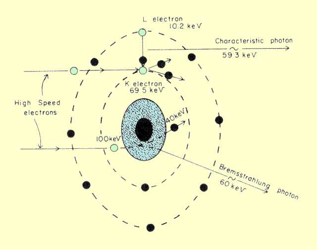
    The type of interaction that produces characteristic radiation, also illustrated above (in the "Kinetic" paragraph), involves a collision between the high-speed electrons and the orbital electrons in the atom. The interaction can occur only if the incoming electron has a kinetic energy greater than the binding energy of the electron within the atom. When this condition exists, and the collision occurs, the electron is dislodged from the atom. When the orbital electron is removed, it leaves a vacancy that is filled by an electron from a higher energy level. As the filling electron moves down to fill the vacancy, it gives up energy emitted in the form of an x-ray photon. This is known as characteristic radiation because the energy of the photon is characteristic of the chemical element that serves as the anode material. In the example shown, the electron dislodges a tungsten K-shell electron, which has a binding energy of 69.5 keV. The vacancy is filled by an electron from the L shell, which has a binding energy of 10.2 keV. The characteristic x-ray photon, therefore, has an energy equal to the energy difference between these two levels, or 59.3 keV.
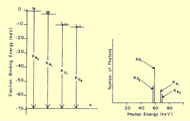
   Actually, a given anode material gives rise to several characteristic x-ray energies. This is because electrons at different energy levels (K, L, etc.) can be dislodged by the bombarding electrons, and the vacancies can be filled from different energy levels. The electronic energy levels in tungsten are shown below, along with some of the energy changes that give rise to characteristic photons. Although filling L-shell vacancies generates photons, their energies are too low for use in diagnostic imaging. Each characteristic energy is given a designation, which indicates the shell in which the vacancy occurred, with a subscript, which shows the origin of the filling electron. A subscript alpha (a) denotes filling with an L-shell electron, and beta ((3) indicates filling from either the M or N shell


   Tungsten Spectrum

   The spectrum of the significant characteristic radiation from tungsten is shown below. Characteristic radiation produces a line spectrum with several discrete energies, whereas Bremsstrahlung produces a continuous spectrum of photon energies over a specific range. The number of photons created at each characteristic energy is different because the probability for filling a K-shell vacancy is different from shell to shell.


Electron Energy Levels in Tungsten and the Associated Characteristic X-Ray Spectrum


   Molybdenum Spectrum

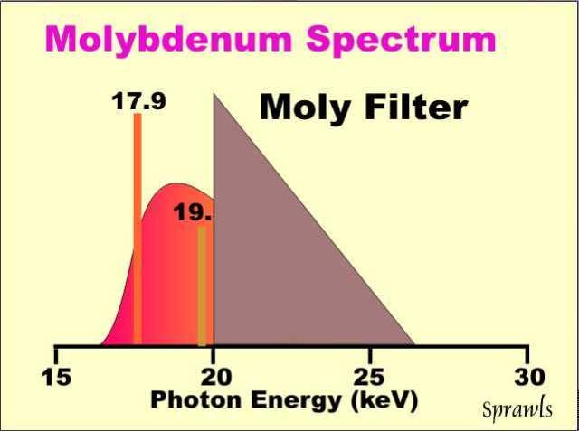
  Molybdenum anode tubes used for mammography produce two rather intense characteristic x-ray energies: K-alpha radiation, at 17.9 keV, and K-beta, at 19.5 keV. as shown below.
The optimum spectrum to produce the best balance between contrast sensitivity and radiation dose for an average size breast is one with most of the radiation with photon energies below about 20 keV.  However, there is considerable Bremsstrahlung above this energy.  In the typical mammography equipment a molybdenum filter is used to remove that undesirable part of the spectrum.  This is an application of a filter that works on the "K edge" principle.  It absorbs radiation that is above the K-edge energy that corresponds to the binding energy of the electrons in the K shell of the molybdenum atom. 

   Rhodium Spectrum

Rhodium has an atomic number (Z) of 45 compared to a Z of 42 for molybdenum. Therefore the characteristic x-radiation produced with a rhodium anode will have energies that are slightly higher than produced with molybdenum and are more penetrating.  This is of value for imaging dense breast.
Anodes that have dual surface areas, molybdenum and rhodium, make it possible for the operator to select a spectrum that is more optimized for different breast sizes and densities.


 KV Effect on Spectrum

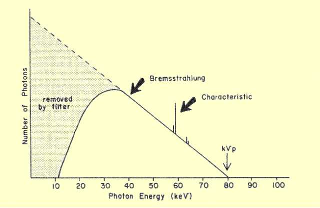
   The KV value also strongly influences the production of characteristic radiation. No characteristic radiation will be produced if the KV is less (numerically) than the binding energy of the K-shell electrons. When the KV is increased above this threshold level, the quantity of characteristic radiation is generally proportional to the difference between the operating KV and the threshold KV.    The x-ray beam that emerges from a tube has a spectrum of photon energies determined by several factors. A typical spectrum is shown below and is made up of photons from both Bremsstrahlung and characteristic interactions.

Typical Photon Energy Spectrum from a Machine Operating at KV = 80

   The relative composition of an x-ray spectrum with respect to Bremsstrahlung and characteristic radiation depends on the anode material, KV, and filtration. In a tungsten anode tube, no characteristic radiation is produced when the KV is less than 69.5. At some higher KV values generally used in diagnostic examinations, the characteristic radiation might contribute as much as 25% of the total radiation. In molybdenum target tubes operated under certain conditions of KV and filtration, the characteristic radiation can be a major part of the total output.

   Concept

    Only a small fraction of the energy delivered to the anode by the electrons is converted into x-radiation; most is absorbed by the anode and converted into heat. The efficiency of x-ray production is defined as the total x-ray energy expressed as a fraction of the total electrical energy imparted to the anode. The two factors that determine production efficiency are the voltage applied to the tube, KV, and the atomic number of the anode, Z. An approximate relationship is
Efficiency = KV x Z x 10-6.

  KV Effect

   The relationship between x-ray production efficiency and KV  has a specific effect on the practical use of x-ray equipment. As we will see in a later chapter, x-ray tubes have a definite limit on the amount of electrical energy they can dissipate because of the heat produced. This, in principle, places a limit on the amount of x-radiation that can be produced by an x-ray tube. By increasing KV, however, the quantity of radiation produced per unit of heat is significantly increased.

   Anode Material

   The relationship of x-ray production efficiency to anode material is only of academic interest because most tubes use tungsten. The exception is molybdenum and rhodium used in mammography. The x-ray production efficiency of these tubes is significantly less than that of tungsten anode tubes because of their lower atomic numbers.

   Definition and Concept

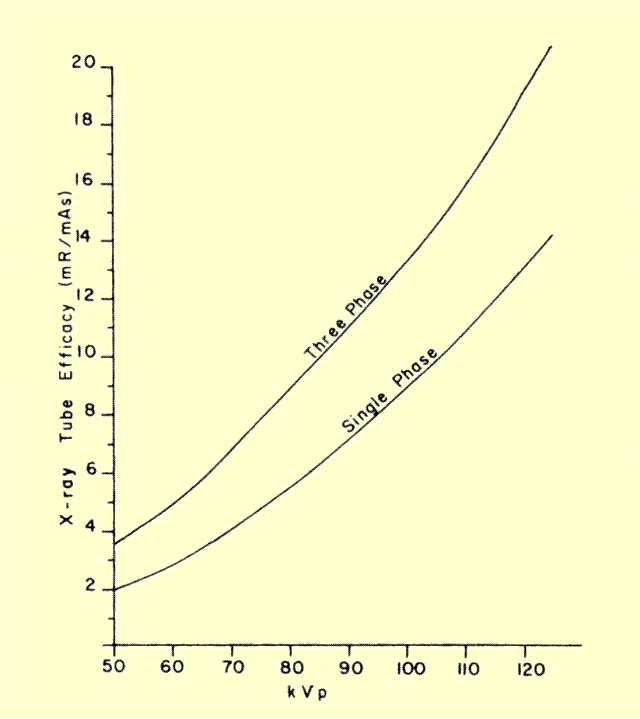
    The x-ray efficacy of the x-ray tube is defined as the amount of exposure, in milliroentgens, delivered to a point in the center of the useful x-ray beam at a distance of 1 m from the focal spot for 1 mAs of electrons passing through the tube.
   The efficacy value expresses the ability of a tube to convert electronic energy into x-ray exposure. Knowledge of the efficacy value for a given tube permits the determination of both patient and image receptor exposures by methods discussed in later chapters. Like x-ray energy output, the efficacy of a tube depends on a number of factors including KV, voltage waveform, anode material, filtration, tube age, and anode surface damage. The illustration below gives typical efficacy values for tungsten anode tubes with normal filtration.

 KV Control

   KV is very useful in controlling the radiation output of an x-ray tube. The figure below shows a nonlinear relationship. It is normally assumed that the radiation output is proportional to the square of the KV. Doubling KV quadruples the exposure from the tube.

Typical X-Ray Tube Efficacy (Exposure Output) for Different KVP Values

   Waveform

   Waveform describes the manner in which the KV changes with time during the x-ray production process because of the cyclic nature of the electrical supply.; several different KV waveforms are used. A general principle is that the waveform with the least KV variation during the exposure is the most effective x-ray producer. Most new x-ray equipment now use  generators that produce relatively constant KV throughout the exposure.  Other waveforms are described in more detail in another chapter. 

   SUMMARY and MIND MAP

The mindmap below provides a summary of the major concepts associated with x-ray production.